Shoulder Anatomy
Before you begin self-massage, it’s a good idea to learn a little about anatomy so you can familiarize yourself with the muscles, bones, and joints of the shoulder and neck.
The Bones
Now let’s look at the bone structure of the shoulder. The shoulder consists primarily of three bones:
- The humerus
- The scapula
- The clavicle
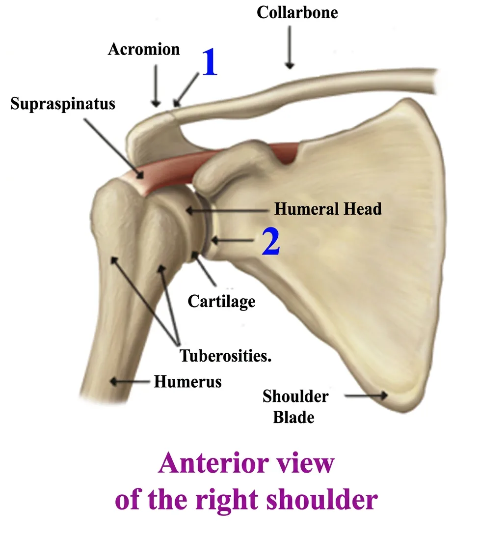
The upper part of the shoulder is formed partly by a projection of the shoulder blade called the acromion.
The Humerus
The humerus is the long bone of the upper arm. The head of the humerus articulates with the glenoid cavity of the scapula. Its lower end articulates with the bones of the forearm, the radius and ulna, to help form the elbow joint.
The Scapula (Shoulder Blade)
The shoulder blade is a flat, triangular-shaped bone located in the upper posterior part of the thorax.
The Clavicle (Collarbone)
From the Latin clavicula, meaning “little key,” the clavicle has an overall shape resembling an inverted “S.” One end connects with the acromion of the scapula at the acromioclavicular joint, while the other connects with the sternum at the sternoclavicular joint.
Shoulder Joints
Pain and stiffness in the shoulder are often attributed to a problem with the glenohumeral joint. You may have heard terms such as inflammation, tendinitis, bursitis, arthritis, or cartilage deterioration.
These conditions can certainly occur. However, muscles and other soft tissues surrounding the shoulder may also contribute significantly to pain, stiffness, and restricted movement.
The glenoid cavity is relatively shallow. This contributes to the tremendous mobility of the shoulder, but it also means the joint depends heavily on the surrounding muscles, tendons, ligaments, and other soft tissues for stability.
Major Joints of the Shoulder
1. The Acromioclavicular Joint
The acromioclavicular joint, commonly called the AC joint, is located where the acromion of the shoulder blade meets the clavicle.
Movement at this joint contributes to the ability of the shoulder blade to move as you raise your arm overhead.
2. The Glenohumeral Joint
Commonly known as the shoulder joint, the glenohumeral joint connects the humerus to the scapula.
The nearly spherical head of the humerus rests in the relatively shallow glenoid cavity of the scapula. This arrangement provides the shoulder with a very large range of motion while requiring considerable muscular and ligamentous support for stability.
3. The Sternoclavicular Joint
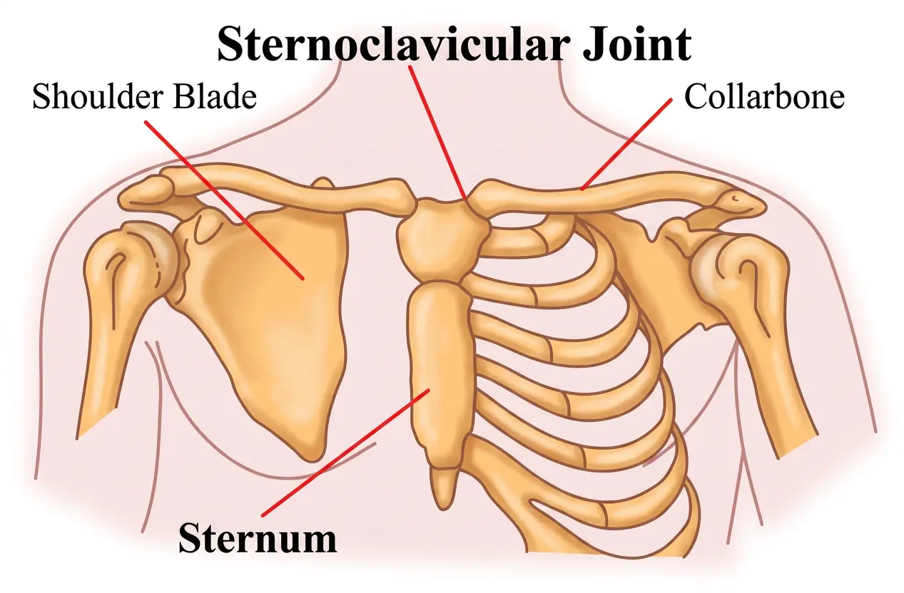
The sternoclavicular joint connects the sternum to the clavicle.
It provides the principal bony connection between the upper limb and the axial skeleton.
4. The Scapulothoracic Joint
The scapulothoracic articulation is not a traditional synovial joint. Instead, it describes the movement of the shoulder blade over the rib cage.
This sliding relationship allows the scapula to move as the arm is raised, lowered, and rotated.
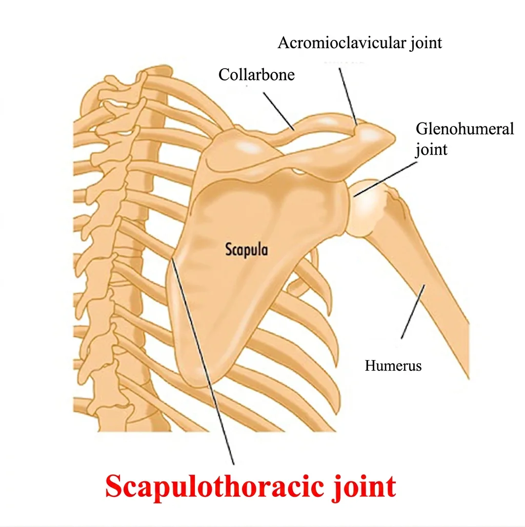
The shoulder complex relies on ligaments, muscles, tendons, and other soft tissues to provide stability while allowing a large range of movement.
Muscles Associated With Shoulder Pain
More than twenty-five muscles can directly or indirectly contribute to pain felt around the shoulder, including muscles located in the neck.
On this website, I organize these muscles according to the general area where discomfort may be felt:
- Muscles that may refer pain to the front of the shoulder
- Muscles that may refer pain to the back of the shoulder
- Muscles that may refer pain to the side of the shoulder
- Muscles that may refer pain to the top of the shoulder
Before looking at all of these muscles individually, I would like to begin with one particularly important group: the muscles of the rotator cuff.
The Rotator Cuff
The rotator cuff is a group of four muscles and their tendons surrounding the shoulder joint. These muscles help stabilize the head of the humerus and contribute to rotation and other movements of the arm.
The four rotator cuff muscles are:
- The Supraspinatus
- The Infraspinatus
- The Teres Minor
- The Subscapularis
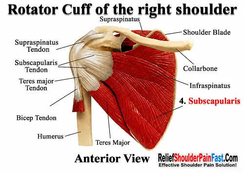
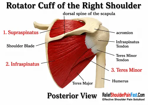
The four muscles of the rotator cuff attach to the shoulder blade and the upper part of the humerus. They perform different movements individually but work together to help stabilize the head of the humerus within the glenoid cavity.
Trigger points and excessive muscular tension in the rotator cuff may contribute to shoulder pain, restricted movement, weakness, and altered shoulder mechanics.
The infraspinatus and subscapularis in particular can develop trigger points that produce pain patterns which may be felt around the shoulder and arm.
Travell and Simons extensively described trigger points in these muscles and their potential relationship with pain and restricted shoulder movement.
Persistent muscular tension can also interfere with normal shoulder mechanics. When pain, weakness, loss of motion, significant clicking, or symptoms persist, an appropriate medical evaluation is advisable.
Reference: Simons, Travell & Simons, 1998, pp. 538–571, 596–607.
Anterior Neck Muscles
Before moving on to the individual shoulder muscles, it is important to remember that some muscles located in the neck can also refer discomfort toward the shoulder.
Two important muscle groups located toward the front and side of the neck are:
- The sternocleidomastoid muscle (commonly abbreviated SCM)
- Scalene muscles
These muscles are located on both sides of the neck. Trigger points and muscular tension within the scalenes may produce referred discomfort into several areas of the shoulder, arm, and upper back.
Thank you for reading,
Michel Bourgeois
Licensed Massage Therapist